microscopy.org
Welcome | Microscopy Society of America
Meeting Minutes and Bylaws. Get Involved in the Society. Microscopy and Microanalysis 2018. M&M Awards and Travel Support. Undergraduate Research Scholarship Program. Standards and Scientific Data. Focused Interest Groups (FIG). The Student Council (StC). MSA Bursary Program M&M 2018. Student Council Media Links. Scholarships, Awards and Recognition. Vote in the MSA Council Elections. Membership Renewal by Invoice. Learn about the latest advances in the field of microscopy. Click here. Are you a member?
 microscopy.org.au
Australian Microscopy and Microanalysis Society – includes the Australian Microbeam Analysis Society (AMAS) and Light Microscopy Australia (LMA)
AMMS is pleased to offer bursaries to our Australian and New Zealand members to assist them in attending the IMC19-ACMM25 conference in Sydney (9-14 September 2018). Membership of AMMS is now online! We invite you to join, renew membership or update your personal details online. Login details have been emailed to current members. 19th International Microscopy Congress (IMC19). International Convention Centre (ICC Sydney). LMA National Meeting 2019. Translational Research Institute Australia, Brisbane.
 microscopy.org.sg
Home
Upcoming Meetings by MS(S). Microscopy centres in Singapore. Workshops by Microscopy Unit. Welcome to the Microscopy Society of Singapore! This is a non-profit think tank actively focused on the promotion of microscopy, organization of microscopy-related activities and facilitation of knowledge and information exchange among its members. MS(S) is moving forward in tandem with new technology and innovations in microscopy, and your input and suggestions for further initiatives are especially welcome.
 microscopy.org.za
:: MWEB Business - Achieve the extraordinary ::
 microscopy.ou.edu
Samuel Roberts Noble Microscopy Laboratory
Samuel Roberts Noble Microscopy Laboratory. Norman, OK 73019-6131. Laboratory, Office and Appointments:. How to get to us:. Forward into the past:. The Samuel Roberts Noble Microscopy Laboratory. SRNEML Personnel and Contact Info:. Preston Larson, Ph.D. Research Scientist, plarson@ou.edu. Tingting Gu, Ph.D. Research Scientist, Confocal Microscopy/Advanced Light Imaging. TEM position is currently vacant. Scott D. Russell, Ph.D. Director, srussell@ou.edu. 8 AM-5 PM M-F. Archive for AME 4143/5143 students.
 microscopy.stanford.edu
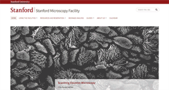
Stanford Microscopy Facility
Access to the Server. Access for non-Stanford, external users. Image Analysis - ImageJ/Fiji. BPAE cells, CSIF. Eva Huang, Dunn Lab, human embryonic stem cells. Confocal microscopy (scanning and spinning-disk). Transmitted-light imaging (phase, DIC, histology). High-content screening (Confocal and Wide-field). Super-resolution imaging (STORM, STED, SIM and AiryScan). Cell surface imaging with 100 nM z-resolution (TIRF). Specialized microscopy (FRAP, FRET, FCS, FLIP.). Chemical and cryo-fixation processing.
 microscopy.synbio.scientific-solution.com
Monkey Patchers
Still in the jungle. But on our way. Please check back after a few bananas! Imprint - Impressum -.
 microscopy.tamu.edu
Welcome to the Microscopy and Imaging Center (MIC) — MIC
Welcome to the Microscopy and Imaging Center (MIC). We serve a wide range of faculty and students at Texas A&M University in addition to researchers from outside of the University. We are promoting cutting edge research in basic and applied sciences through research and development activities, as well as quality training and education. Through individual training, short courses and formal courses that can be taken for credit. Picture of the Month. Click on the image to learn more. Selected labs in the MI...
 microscopy.tll.org.sg
Bioimaging and Biocomputing Facility | TLL
Objectives and Filter Sets. Request for HPC Account. TLL's Galaxy and UCSCGB. Image of the Month. Invasive hyphal growth of Magnaporthe oryzae in live rice cells. Conidia of the M. oryzae were incubated on the rice sheath. Two days after inoculation, the fungal hyphae invaded a neighboring rice cell through plasmodesmata (arrows). The image was taken on the spinning disk confocal, acquired with 60X/1.4 objective. Taken by: Yang Fan.
 microscopy.uark.edu
Arkansas Nano & Bio Materials Characterization Facility | nanoscale instruments & expertise for campus & the state
Arkansas Nano and Bio Materials Characterization Facility. Nanoscale instruments and expertise for campus and the state. Dr Mourad Benamara,. Office: 479.575.7634. Titan’s lab: 479-575-7642. Institute for Nanoscience & Engineering. Dr Gregory Salamo,. 2015, The University of Arkansas.
 microscopy.ucsd.edu
Microscopy Core - UC San Diego
Please note that links from this site point to an updated Microscopy Core Website. For information about the UCSD Cancer Center's Shared Microscopy Resource please click here. The UCSD Health Sciences Microscopy Core is a state-of-the-art imaging core facility that serves the needs of laboratories in and outside of the UCSD School of Medicine. The Core strives to promote interdisciplinary, collaborative research among the local research community. Cancer Center Imaging Resources. UCSD School of Medicine.